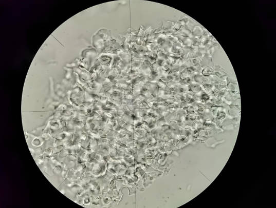
Cell Mechanics Experiment – Accredited Single‑Cell and Monolayer Mechanical Property Evaluation for Global Markets
Our internationally accredited laboratory provides a specialist cell mechanics experiment service that enables pharmaceutical developers, biomedical researchers, tissue‑engineering companies, cancer‑biology teams and medical‑device manufacturers worldwide to independently quantify the viscoelastic, adhesive and force‑generation properties of individual living cells and confluent cell monolayers. Every measurement is performed under the rigorous framework of ISO/IEC 17025, and each report bearing the ILAC mark is unconditionally accepted by regulatory authorities, notified bodies, research funding agencies and supply‑chain partners in all major economies. The cell mechanics experiment platform employs atomic force microscopy, micropipette aspiration, parallel‑plate rheometry, traction‑force microscopy and magnetic twisting cytometry to measure the key mechanical parameters – the Young's modulus, the cortical tension, the viscoelastic creep compliance, the storage and the loss moduli, the adhesion force and the traction stress – that govern the behaviour of the cell during the migration, the division, the differentiation and the malignant transformation. For a biopharmaceutical company screening a cytoskeleton‑targeting anti‑cancer compound, a tissue‑engineering start‑up characterising the stiffness of a stem‑cell‑derived cardiomyocyte sheet, or a medical‑device producer evaluating the effect of a novel stent coating on the endothelial‑cell mechanics, this service delivers the legally robust, defensible biophysical data that underpin the drug‑development pipeline, the regulatory submission and the publication in high‑impact scientific journals.
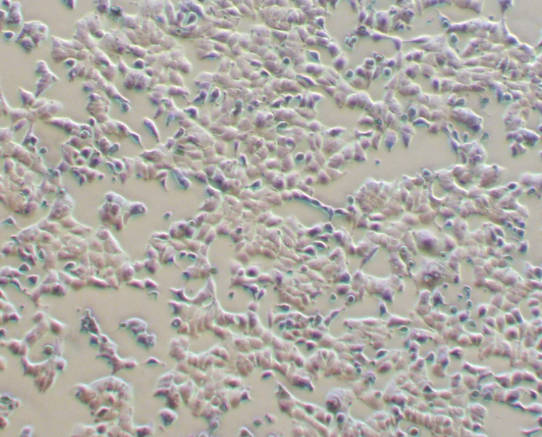
Product Samples We Regularly Subject to the Cell Mechanics Experiment
The atomic‑force‑microscope, the micropipette‑aspiration rig, the parallel‑plate rheometer, the inverted‑fluorescence‑microscope traction‑force workstation and the magnetic‑twisting‑cytometry system in our facility accommodate a wide variety of cell types and culture formats. The following categories represent the most frequently tested items:
- Mammalian cell lines and primary cells – HeLa, NIH/3T3, MCF‑7, MDA‑MB‑231, HEK‑293, human umbilical vein endothelial cells, human dermal fibroblasts, mesenchymal stem cells and the induced pluripotent stem‑cell‑derived lineages, cultured as the single cells or the confluent monolayers
- Circulating and the blood‑lineage cells – the erythrocytes, the leukocytes, the lymphocytes, the monocytes and the platelets, evaluated for the deformability, the aggregation and the adhesion properties that are critical for the haemorheology and the immune‑cell trafficking
- Cancer and the tumour‑derived cells – the primary tumour cells, the patient‑derived xenograft cells and the circulating tumour cells, characterised for the stiffness, the contractility and the invasive‑migration force that correlate with the metastatic potential
- Stem‑cell and the organoid cultures – the embryonic stem cells, the induced pluripotent stem‑cell colonies and the intestinal, the hepatic and the cerebral organoids, measured for the regional mechanical heterogeneity that directs the lineage specification and the morphogenesis
- Pharmacologically and the genetically modified cells – the cells that have been treated with the cytoskeleton‑targeting drugs, the gene‑silencing RNA, the CRISPR‑edited knock‑outs or the substrate‑stiffness‑conditioned media, submitted for the quantification of the treatment‑induced mechanical phenotype
- Bacteria, yeast and the plant cells – the Escherichia coli, the Saccharomyces cerevisiae and the Arabidopsis thaliana protoplasts, evaluated for the turgor pressure, the cell‑wall elasticity and the osmotic‑stress response
Atomic Force Microscopy – Nano‑Indentation and Force‑Spectroscopy Cell Mechanics Experiment
- Determination of the apparent Young's modulus of the single cell by the colloidal‑probe atomic‑force‑microscopy nano‑indentation according to the internal validated protocols and the best‑practice guidelines of the European Network on Cell Mechanics: a tipless cantilever carrying a borosilicate‑glass or a polystyrene microsphere of a defined diameter is brought into the contact with the apical surface of the adherent cell, and the force‑indentation curve is recorded at a controlled approach and the retraction speed. The Hertz or the Sneddon model, corrected for the finite cell thickness and the substrate effect, is fitted to the loading curve, and the apparent Young's modulus in pascals is reported. This cell mechanics experiment provides the most widely used, high‑spatial‑resolution stiffness measurement that distinguishes the benign from the malignant cells, the quiescent from the activated fibroblasts, and the differentiated from the undifferentiated stem cells.
- Force‑spectroscopy mapping and the stiffness‑distribution histogram: an array of the indentation points – typically a 10 × 10 or a 20 × 20 grid covering the entire cell body – is programmed, and the Young's modulus is calculated at each point, generating the two‑dimensional stiffness map and the histogram of the modulus distribution that reveals the sub‑cellular mechanical heterogeneity associated with the actin‑stress‑fibre bundles, the focal adhesions and the nucleus.
- Measurement of the single‑cell adhesion force and the work of the adhesion by the retraction‑curve analysis: after a defined contact dwell‑time, the cantilever is retracted, and the maximum force required to detach the probe from the cell surface, and the area under the retraction curve (the work of the adhesion), are reported, quantifying the strength of the cell‑surface receptor–ligand bonds and the effect of the adhesion‑enhancing or the adhesion‑blocking drugs.
- Viscoelastic characterisation by the stress‑relaxation and the oscillatory‑indentation methods: the indenter is held at a constant depth, and the decay of the force with the time is recorded, or the sample is indented with a small‑amplitude sinusoidal modulation, and the storage and the loss moduli and the relaxation time constant are extracted, providing the time‑dependent mechanical parameters that reflect the fluid‑like viscous behaviour of the cytoplasm and the solid‑like elastic behaviour of the actin cortex.
Micropipette Aspiration – Cortical Tension and the Whole‑Cell Viscoelasticity in the Cell Mechanics Experiment
- Determination of the cortical tension and the apparent viscosity of the single suspended cell according to the micropipette‑aspiration method based on the principles of Evans and Yeung and the internal protocols: a single cell is aspirated into a glass micropipette with an inner diameter slightly smaller than the cell, and the aspiration pressure is increased in the controlled steps. The length of the cell tongue inside the pipette as a function of the applied pressure is recorded, and the cortical tension in newtons per metre and the apparent viscosity in pascal‑seconds are calculated from the linear‑regression slope and the creep‑recovery kinetics. This cell mechanics experiment is the classical biophysical method for the characterisation of the erythrocyte deformability, the leukocyte stiffness and the oocyte cortical tension, and it provides the parameters that are directly comparable with the extensive published literature.
- Step‑pressure creep and the recovery testing for the full viscoelastic characterisation: a constant aspiration pressure is applied for a defined period, and the time‑dependent elongation of the cell tongue is recorded (the creep phase), after which the pressure is released and the retraction of the tongue is monitored (the recovery phase). The creep‑compliance curve is analysed with the generalised Maxwell‑Wiechert or the power‑law rheology model, and the spectrum of the relaxation times is reported, providing the comprehensive viscoelastic fingerprint of the cell.
- Measurement of the cell‑cell adhesion force by the dual‑micropipette method: two cells are held in two separate micropipettes, brought into the controlled contact for a defined time, and then pulled apart while the force required to separate them is calculated from the deflection of the calibrated micropipette, quantifying the strength of the cadherin‑mediated intercellular adhesion that is critical for the tissue integrity and the cancer metastasis.
Traction‑Force Microscopy – Quantifying the Cell‑Generated Substrate Stresses
- Determination of the cell‑generated traction stress by the hydrogel‑substrate traction‑force microscopy according to the internal validated procedures: the cells are cultured on a compliant polyacrylamide or a polydimethylsiloxane hydrogel substrate that is embedded with the fluorescent micro‑beads. The displacement of the beads caused by the cell‑exerted forces is measured by comparing the image of the stressed substrate with the image of the same field after the cell is detached. The traction‑stress field in pascals is reconstructed from the bead‑displacement field using the Fourier‑transform or the finite‑element method, and the total strain energy and the maximum traction stress are reported. This cell mechanics experiment directly measures the physical forces that the cell applies to its environment, which are the key drivers of the wound‑healing, the tissue morphogenesis, the cancer invasion and the stem‑cell differentiation.
- Time‑lapse traction‑force microscopy and the force‑dynamics analysis: the fluorescent‑bead images are acquired at the 1‑ to 5‑minute intervals for several hours, and the evolution of the traction‑stress field, the force‑dipole moment and the cell‑migration velocity are analysed, revealing the cyclic force‑generation pattern, the force‑transmission dynamics and the correlation between the traction stress and the cell‑shape change during the migration and the division.
- High‑throughput traction‑force screening using the micro‑patterned substrates: the cells are plated on the arrays of the micro‑patterned adhesive islands of a defined shape and area, and the average traction stress and the force‑distribution pattern are measured for the hundreds of the single cells per condition, enabling the statistically powered comparison of the drug treatments, the genetic perturbations and the substrate‑stiffness effects on the cell contractility.
Report Acceptance and Global Regulatory Compliance
All measurements performed within our cell mechanics experiment programme are executed under the fully accredited scope of our ISO/IEC 17025 quality management system. Each test report that carries the ILAC mark is therefore automatically recognised by regulatory authorities, notified bodies, research funding agencies and supply‑chain partners in all major economies. For pharmaceutical developers, biomedical‑research organisations, tissue‑engineering companies and medical‑device manufacturers anywhere in the world, the report constitutes legally robust, internationally accepted evidence that the Young's modulus, the cortical tension, the viscoelastic parameters, the adhesion force and the traction‑stress field of the living cell have been determined in accordance with the internal validated protocols and the customer‑specified methods. The documentation can be directly used to support the investigational‑new‑drug application, the regulatory submission for the medical device, the publication in the peer‑reviewed journal, the issue of inspection certificates according to EN 10204 or equivalent national standards, and the resolution of commercial and technical disputes concerning the biophysical properties and the mechanical phenotype of any eukaryotic or prokaryotic cell.